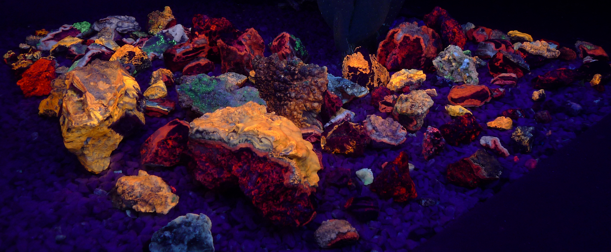
Fluorescent Minerals from Southern Arizona
Latest update:
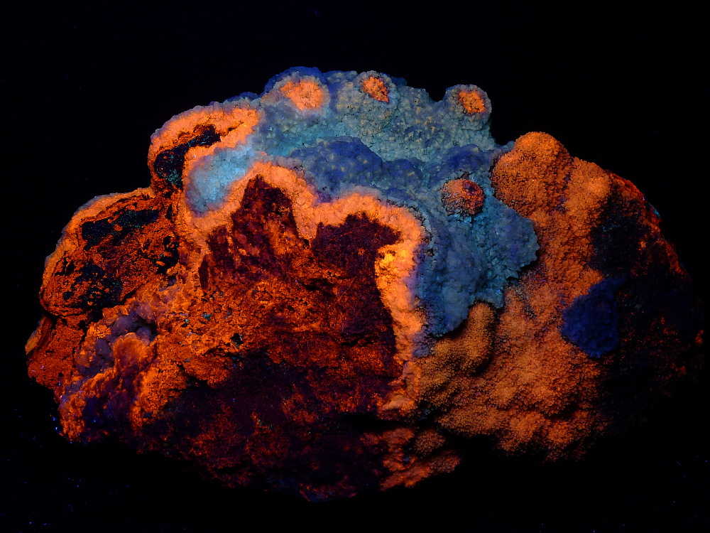
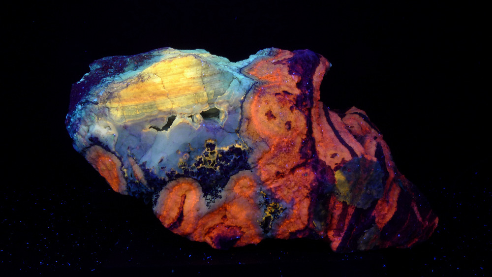
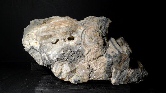
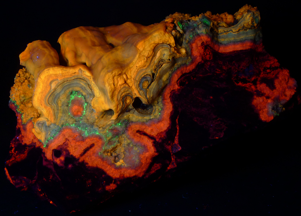
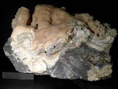
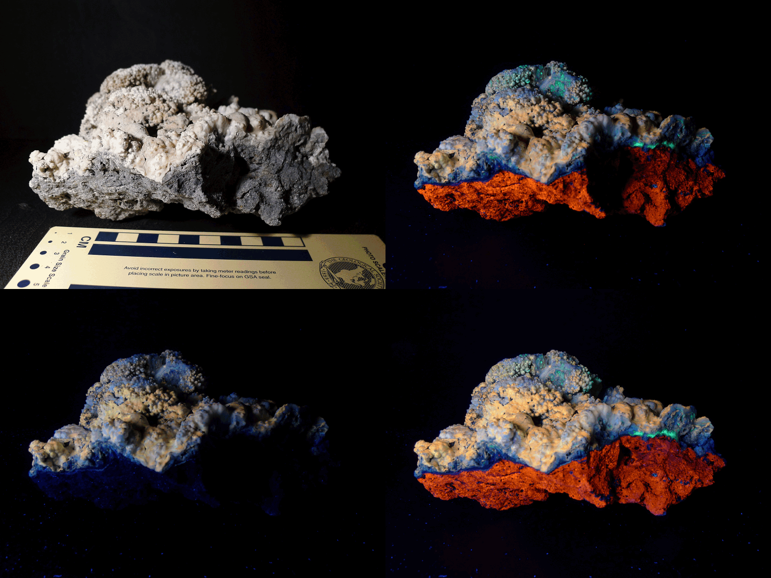
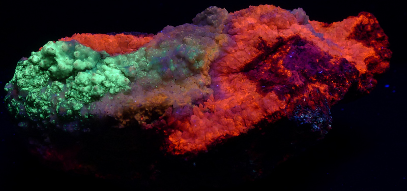
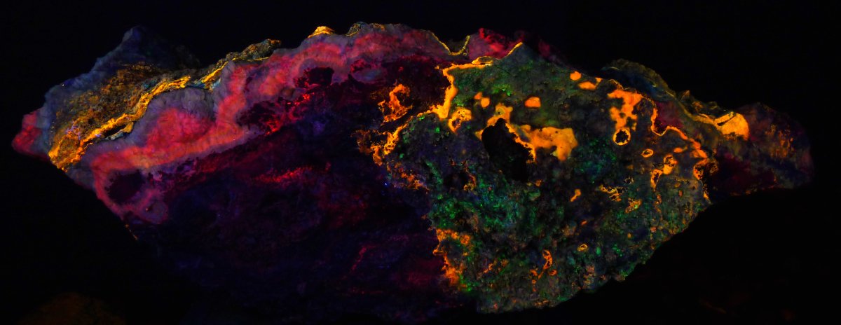
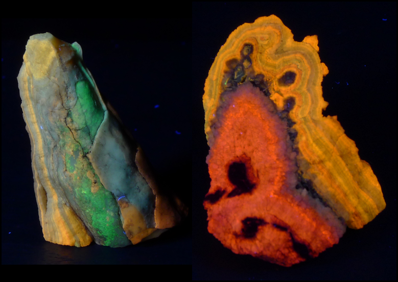
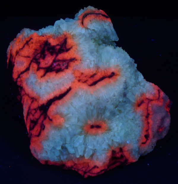
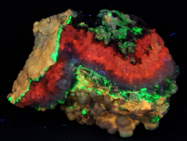
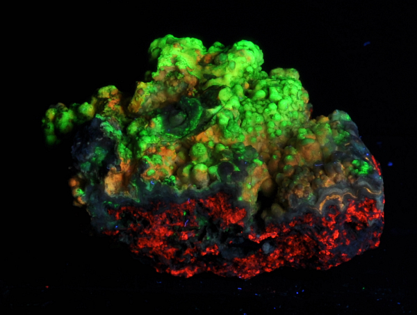
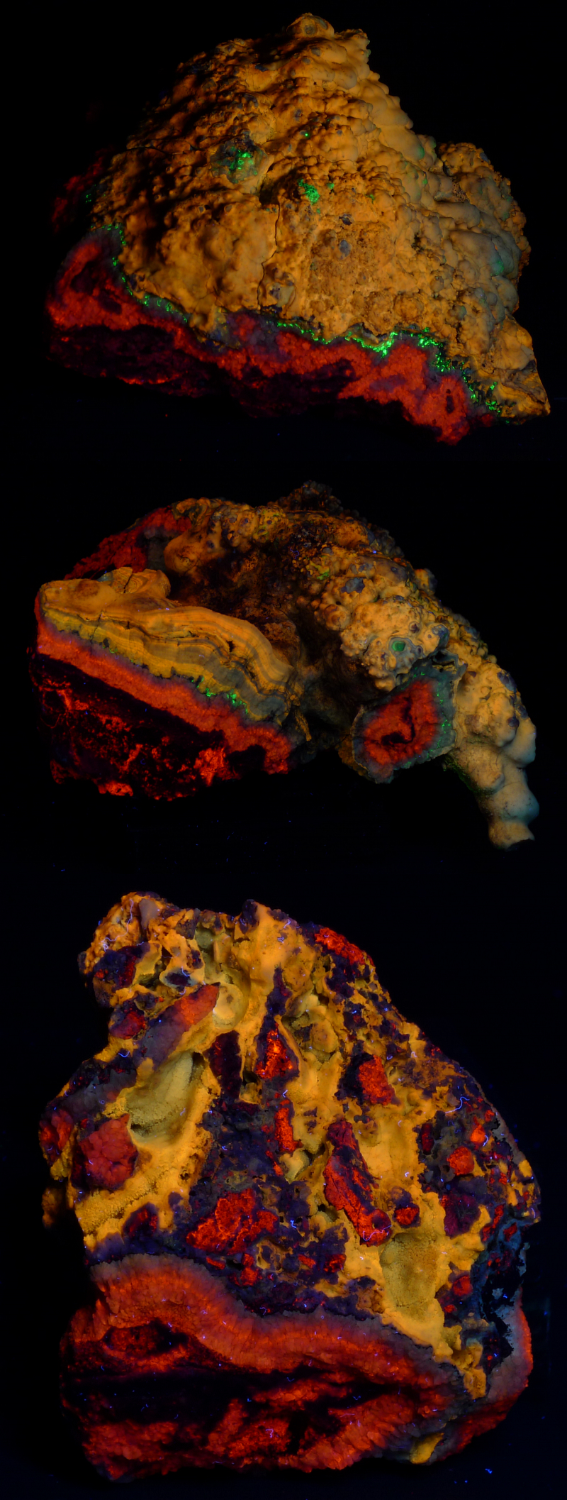
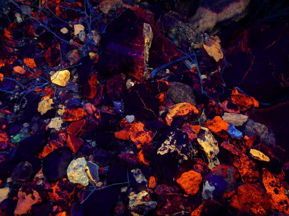
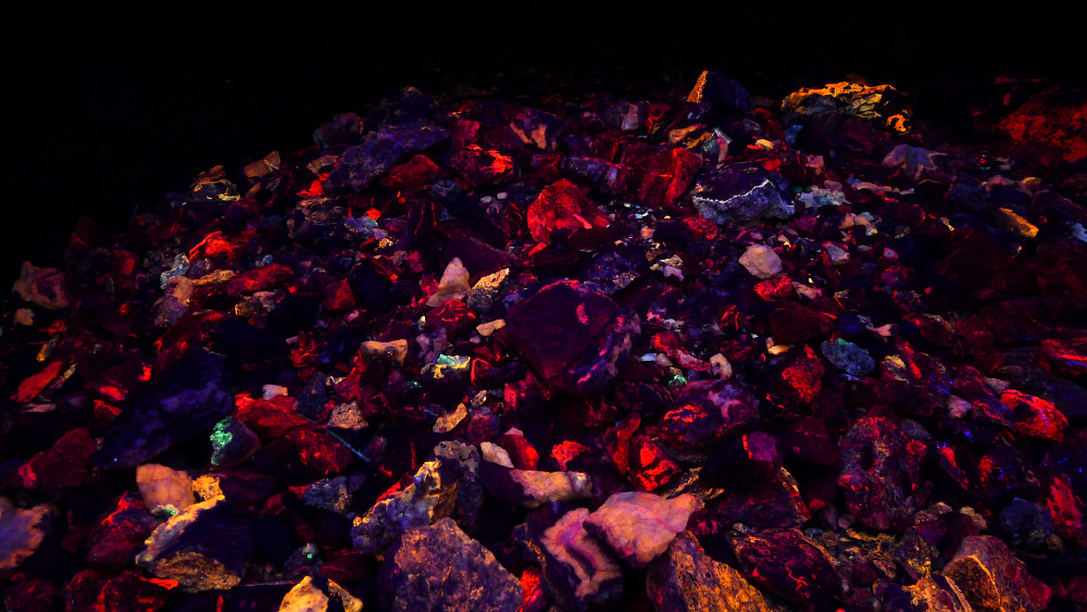
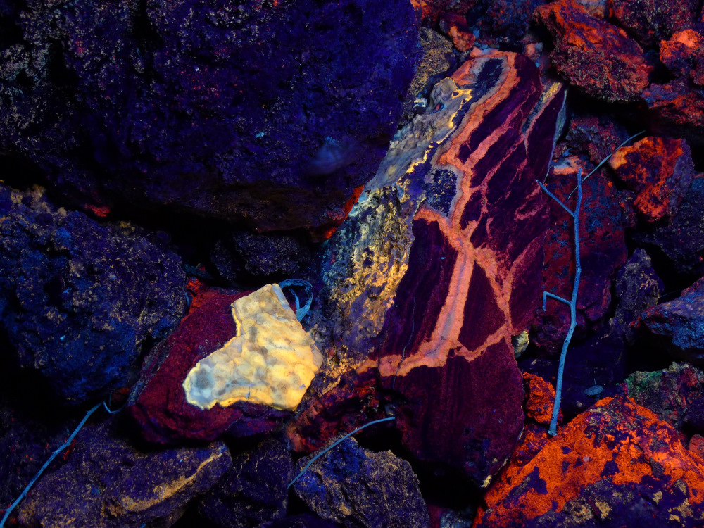
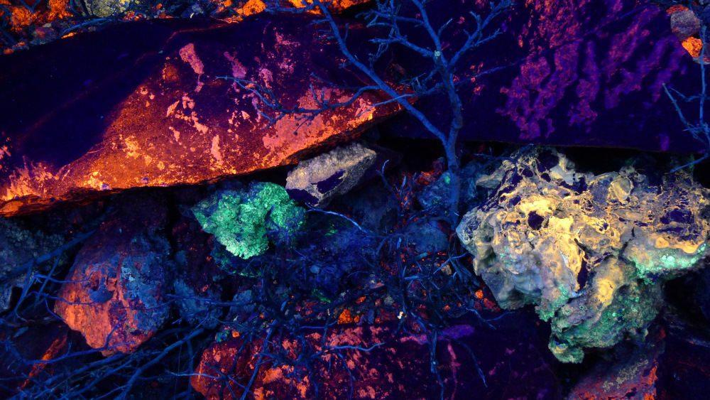
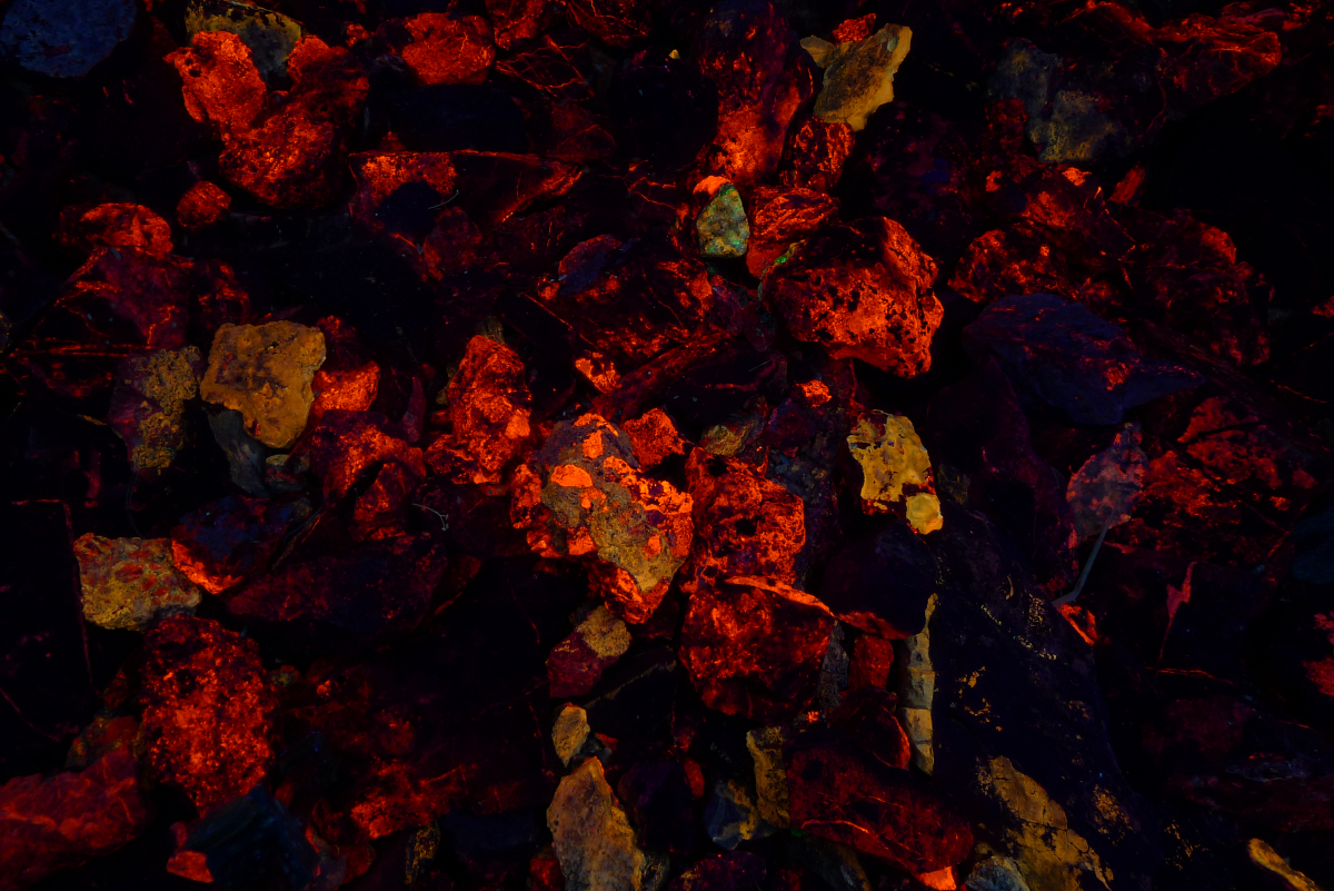
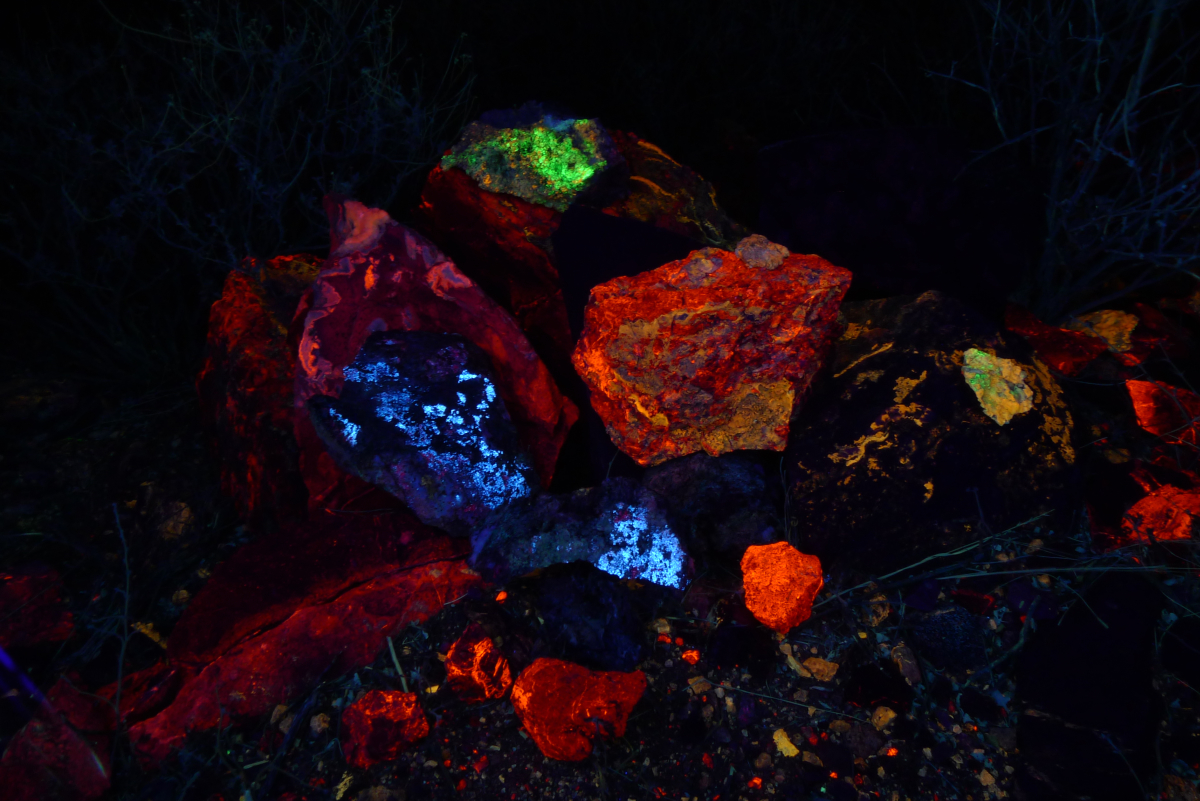
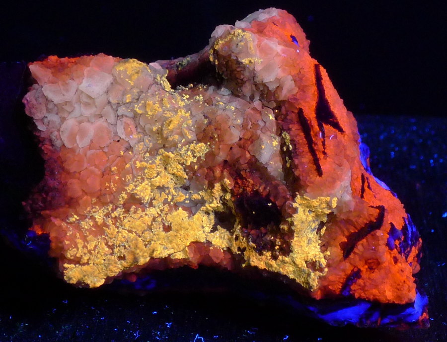
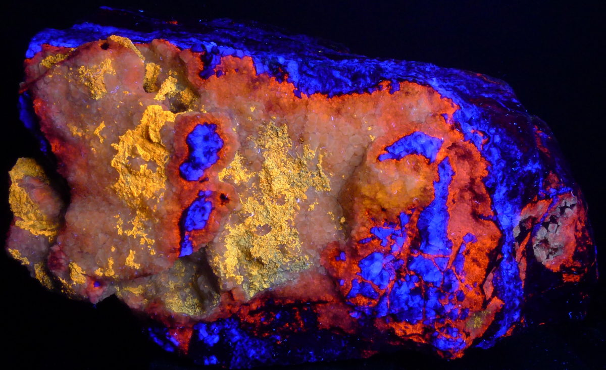
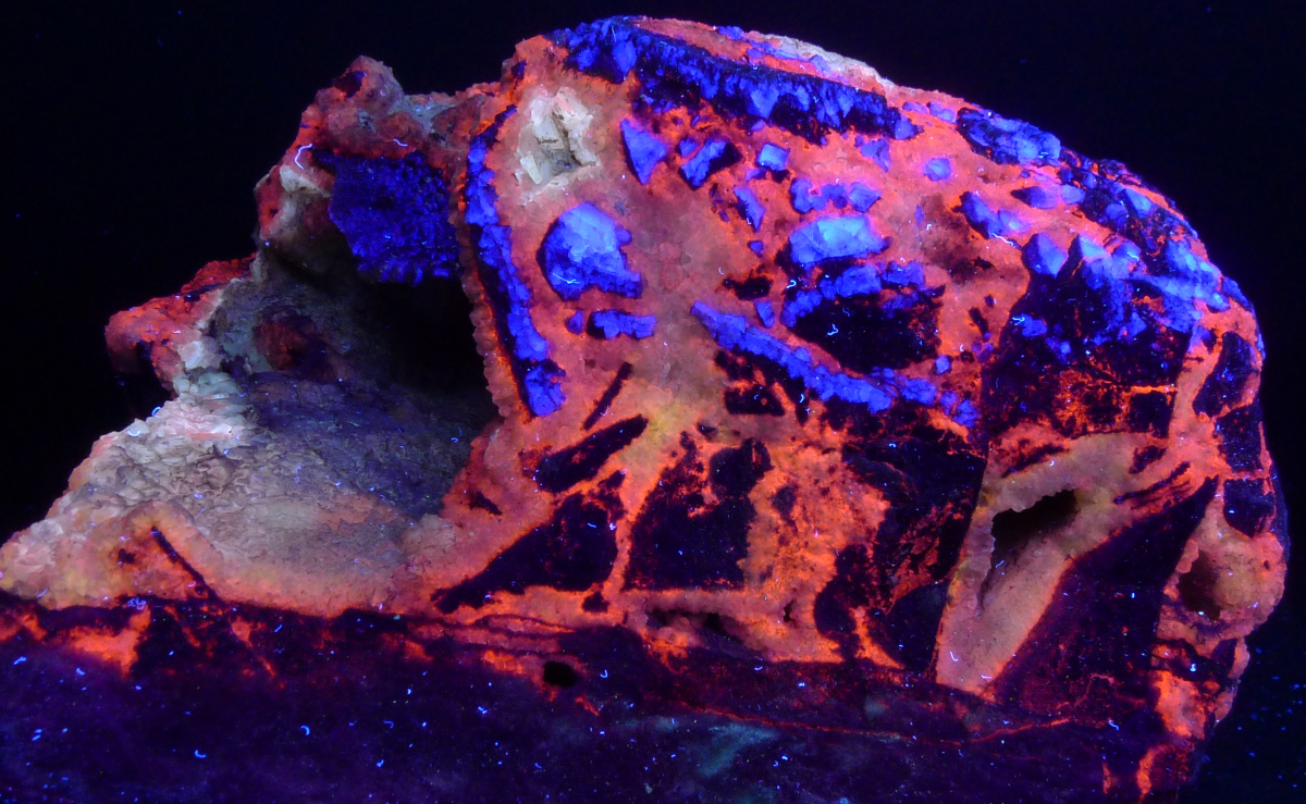
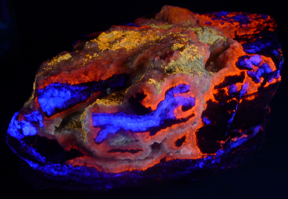
The fluorescent rocks displayed below were collected from a single mine in the mountains of southern Arizona. The rocks consist of calcite, aragonite, and barite with minor amounts of opal, wulfenite, and amphiboles. The fluorescence is the result of minor impurities in the minerals slightly altering chemical bonds in the crystal structures of the minerals.
The Paleozoic limestone host rock was heated and brecciated as a result of the nearby intrusion of magma. Tremolite and muscovite replaced silty layers in the limestone. At least two episodes or pulses of hydrothermal fluids passed through the limestone, one of which carried the trace element(s) responsible for the fluorescence. The hydrothermal alteration of the limestone resulted in the dissolution of portions of the limestone and deposition of calcite veins, overgrowths, and cavity fillings in the rock. A thin layer of hyalite opal was deposited over the calcite. The limestone was later chemically weathered, again leading to dissolution of the rock and the deposition of aragonite (travertine) in dissolution cavities.
| Mineral List (by relative abundance) | |
|---|---|
|
|
| S = shortwave, L= longwave, P = phosphorescent | |
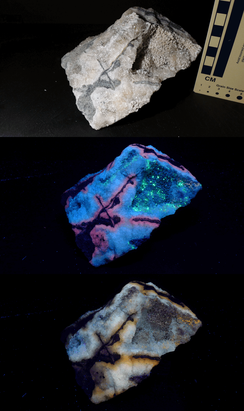
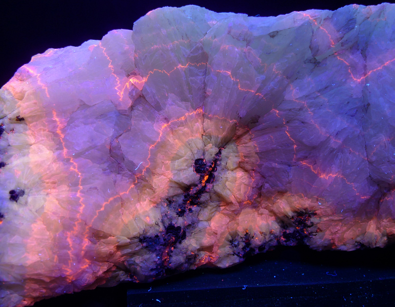
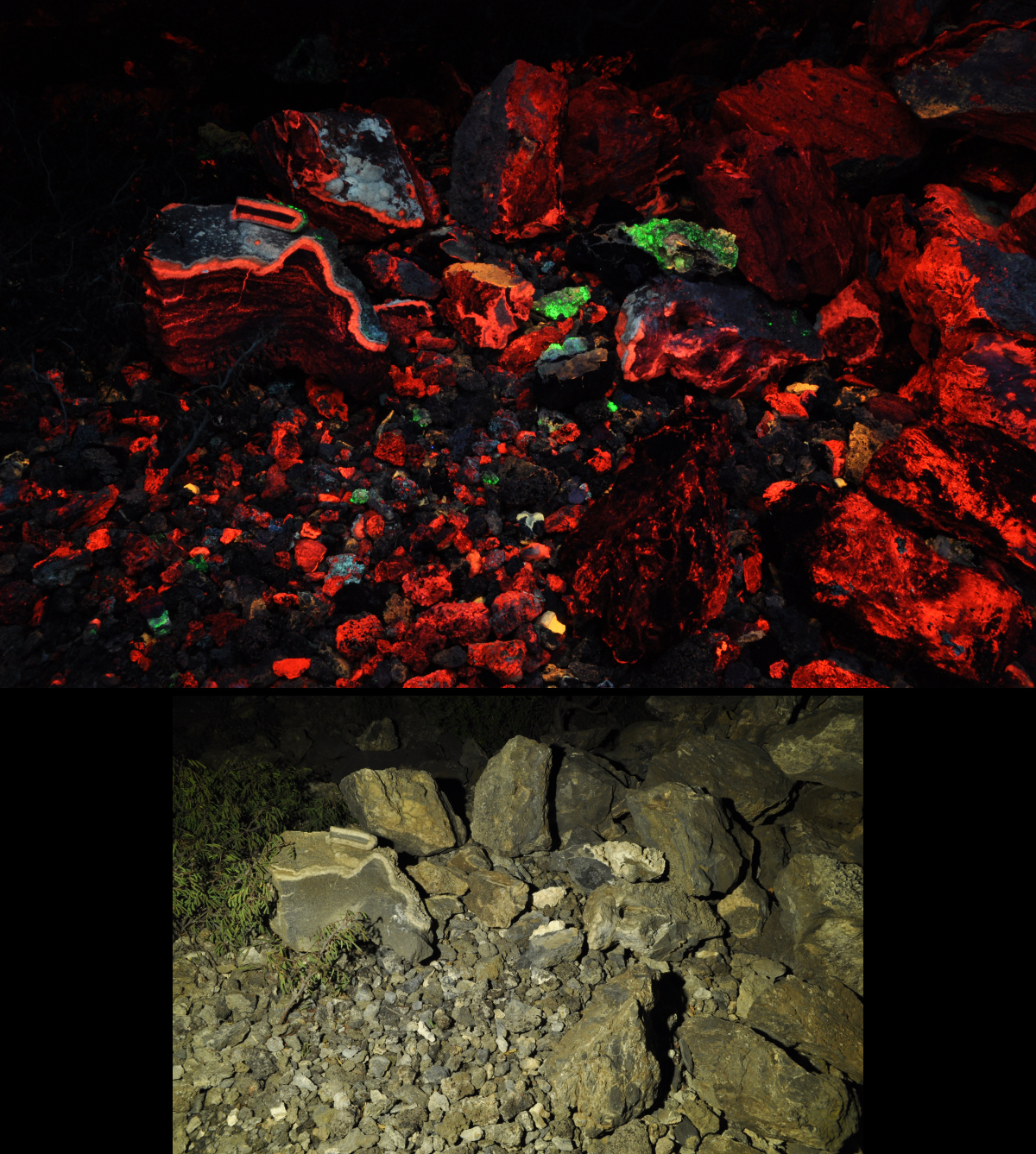
The rocks were photographed under shortwave and longwave ultraviolet light. Unless noted, any blue fluorescence showing in the photos is an artifact of the camera sensor and is white or gray to the naked eye. Bright blue specs are dust particles.
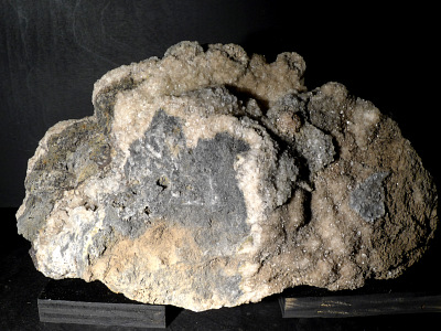
Mouse over the images to read the descriptions. Maximum dimensions given in centimeters.
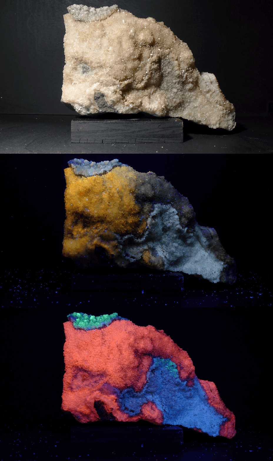
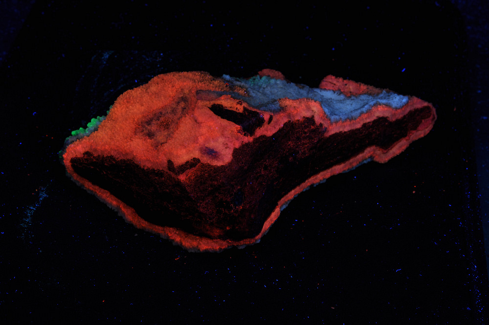
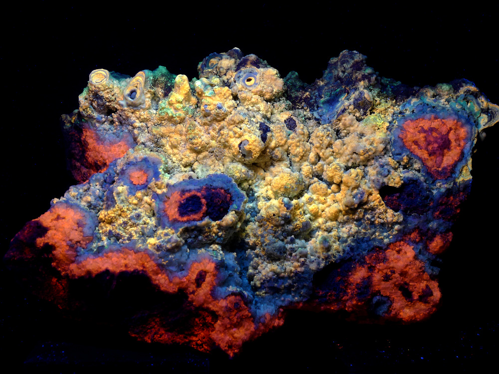
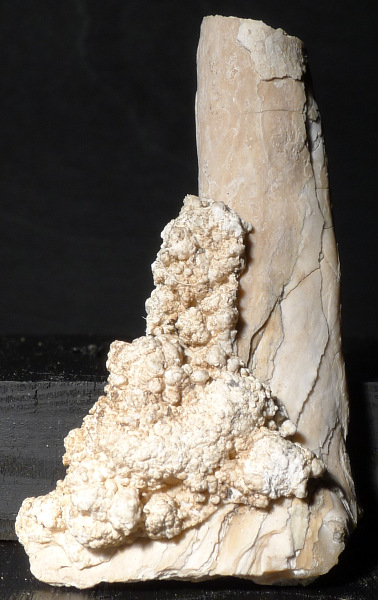
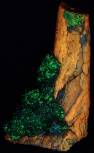
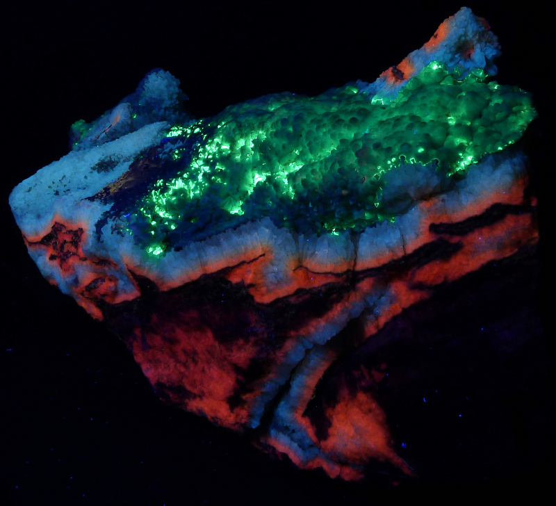
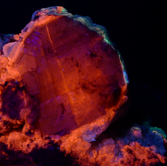
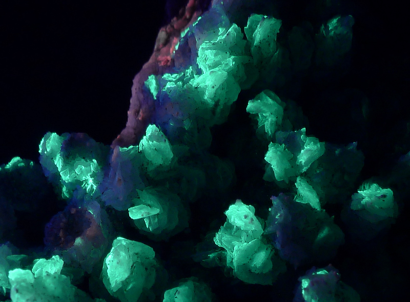
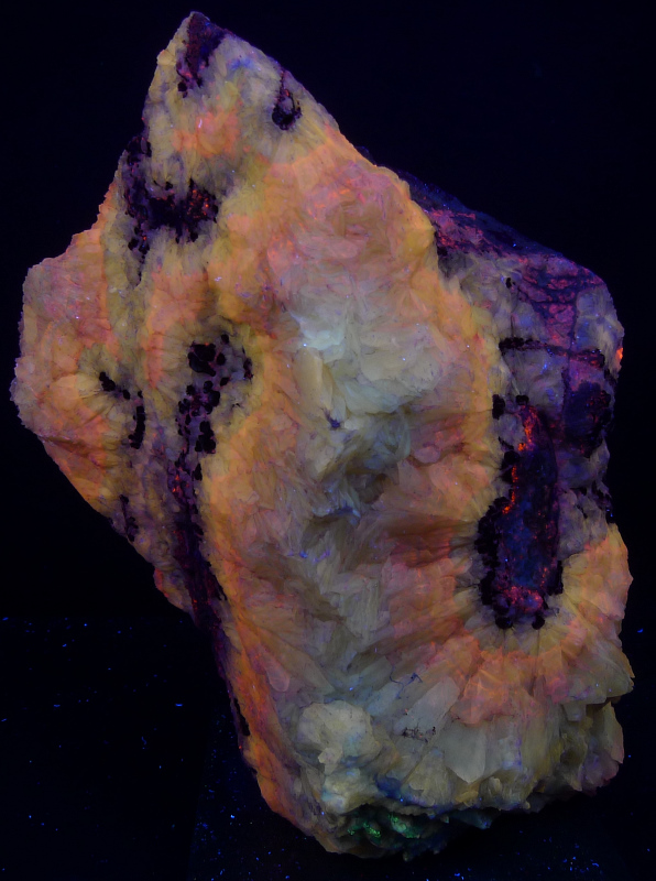
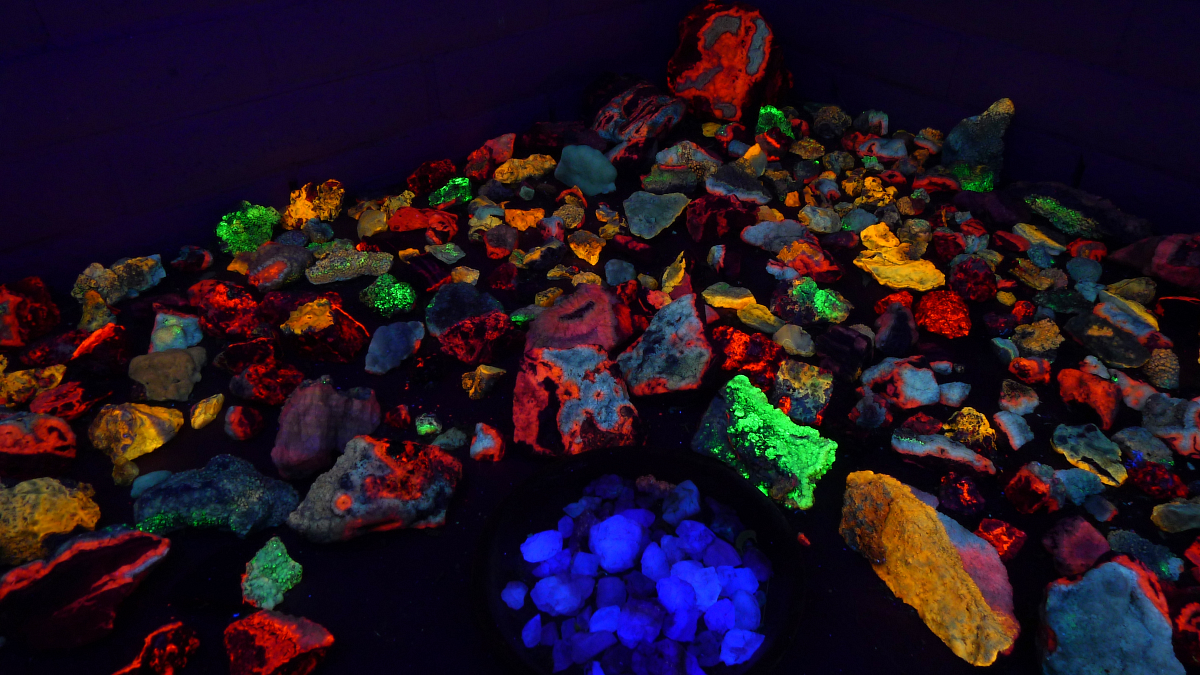
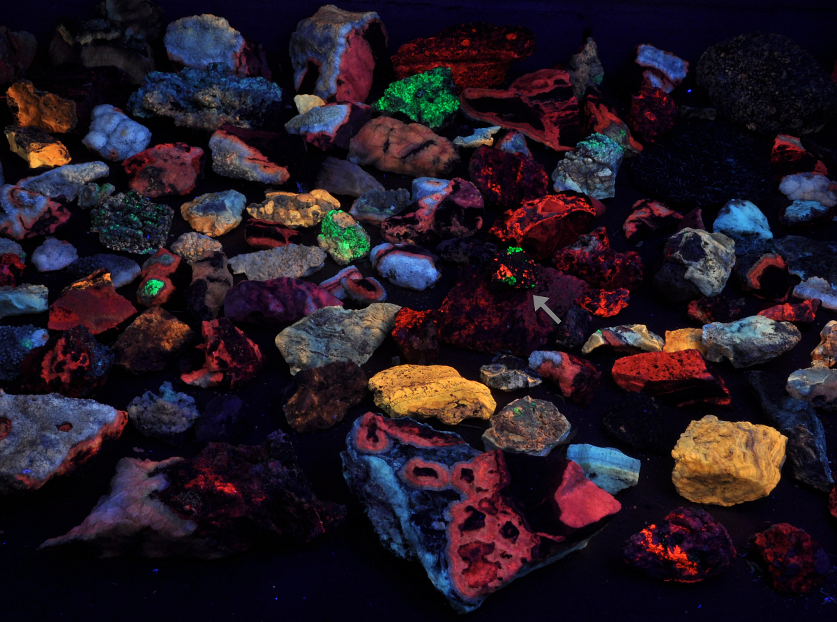
This photo compares the fluorescence of the Arizona specimens to a small specimen from Franklin, NJ under shortwave light. The gray arrow points to the Franklin specimen which shows orange calcite and green willemite. The Franklin specimen is about five centimeters wide.
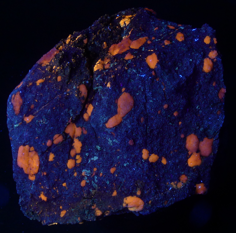
Orange fluorescing gypsum in altered limestone. Fine crystals of tremolite cause the limestone matrix to fluoresce.
Petrographic Thin Sections
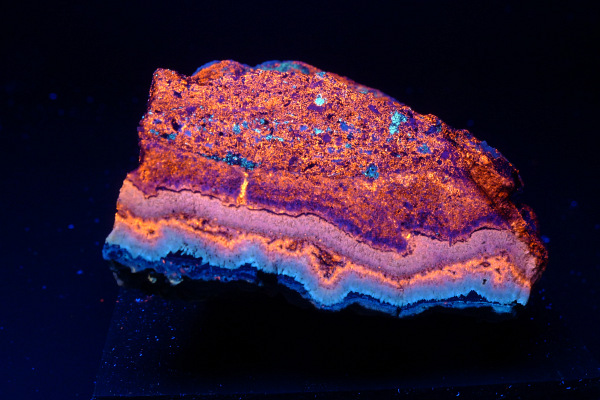
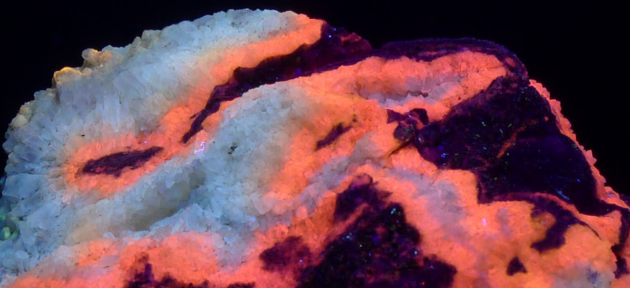
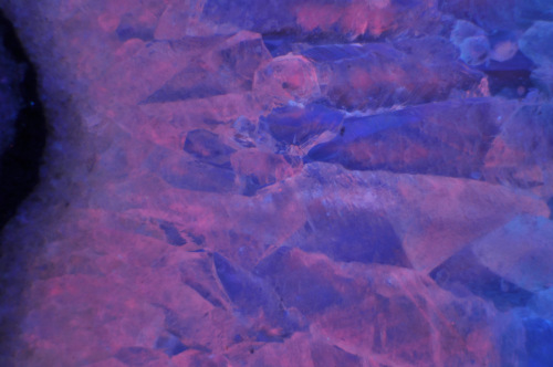
Below are views of a petrographic thin section, ~60 microns in thickness, of the above rock. We are preparing more thin sections and will add them to this page as they are completed.
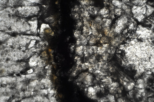
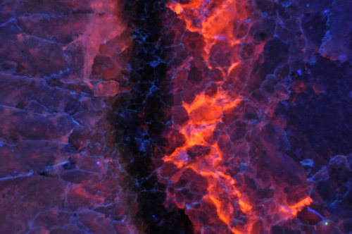
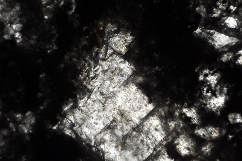
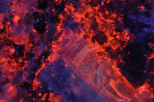
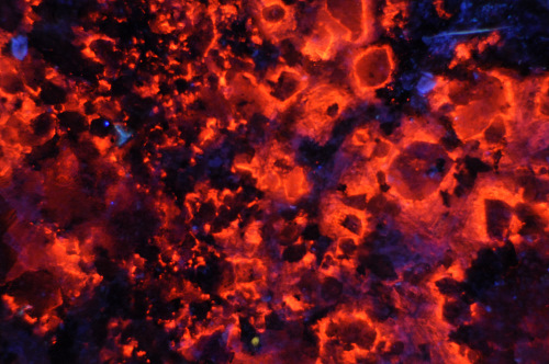
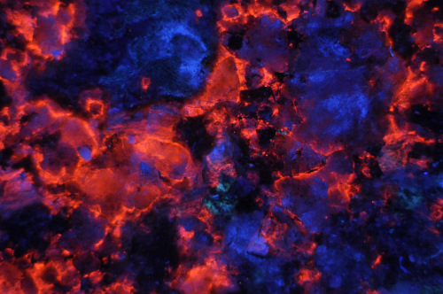
Below is a thin section of a sample taken from a small prospect near the mine. These rocks show a "bleeding" fluorescence forming halos around the host limestone. The white calcite is highly phosphorescent while the orange calcite shows no phosphorescence
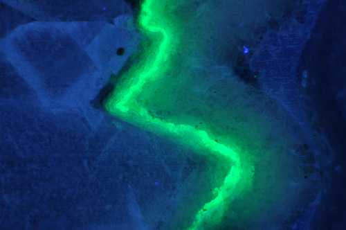
Below is a thin section of a sample showing green fluorescing opal in calcite. Note the difference in calcite crystal habit on either side of opal.
Pictures from the Mine
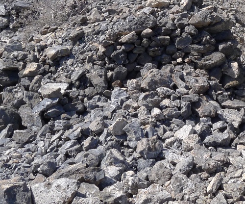
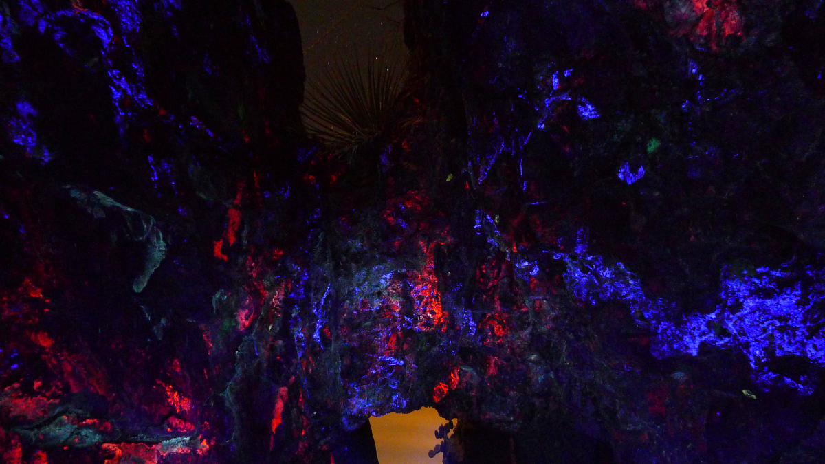
The source mine was worked for metals in the late 19th through early 20th century. The mine dump is the source of all the rocks above.
The bluish white fluorescing mineral below is hydrozincite on sphalerite, a first for this location.
Other Locations Around Southern Arizona
Miller Canyon, Huachuca Mountains, AZ
The specimens below come from a zinc deposit in Miller Canyon in the Huachuca Mountains of Arizona. This is a well known locality for fluorescent minerals.
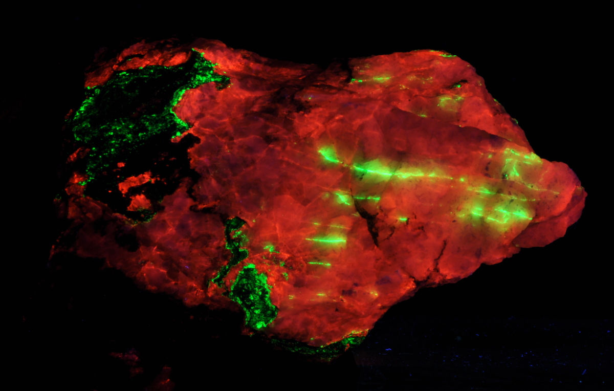
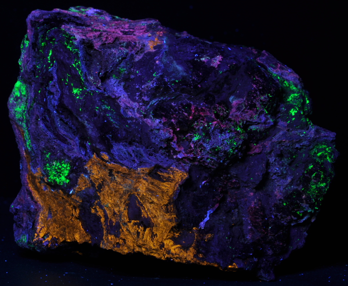
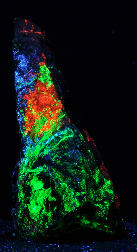
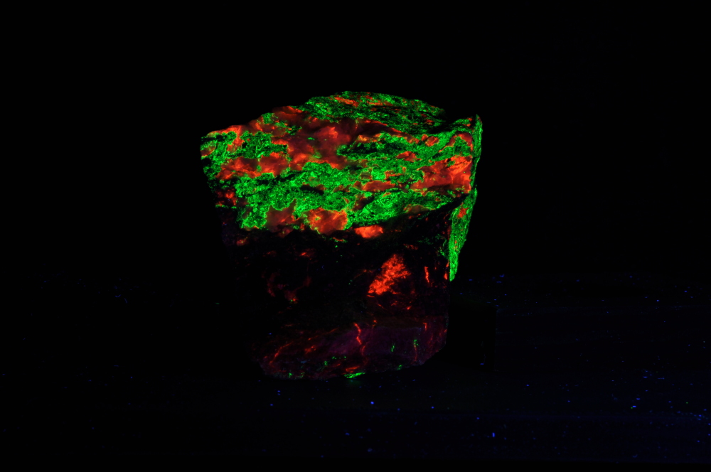
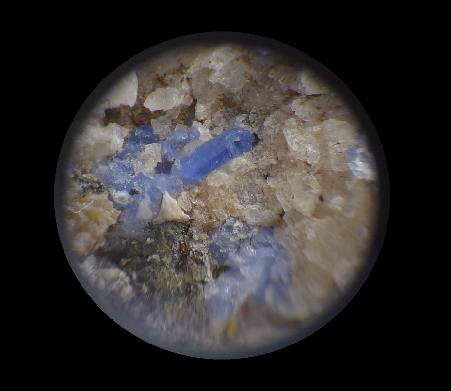
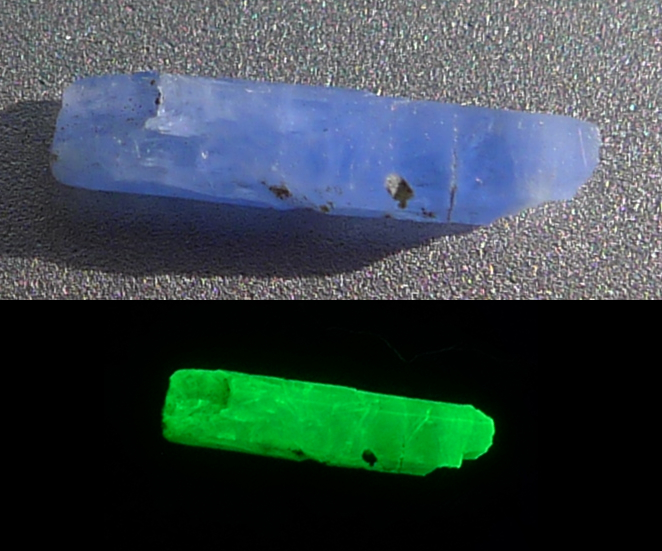
A rarity from Miller Canyon is blue willemite. The blue willemite from Miller Canyon tends to form compact crystals and fluoresce more strongly and phorphoresce longer than the more common willemite coatings shown in the specimens above. The cause of the blue coloring is currently undetermined.
Fluorescent Quartz Crystals!
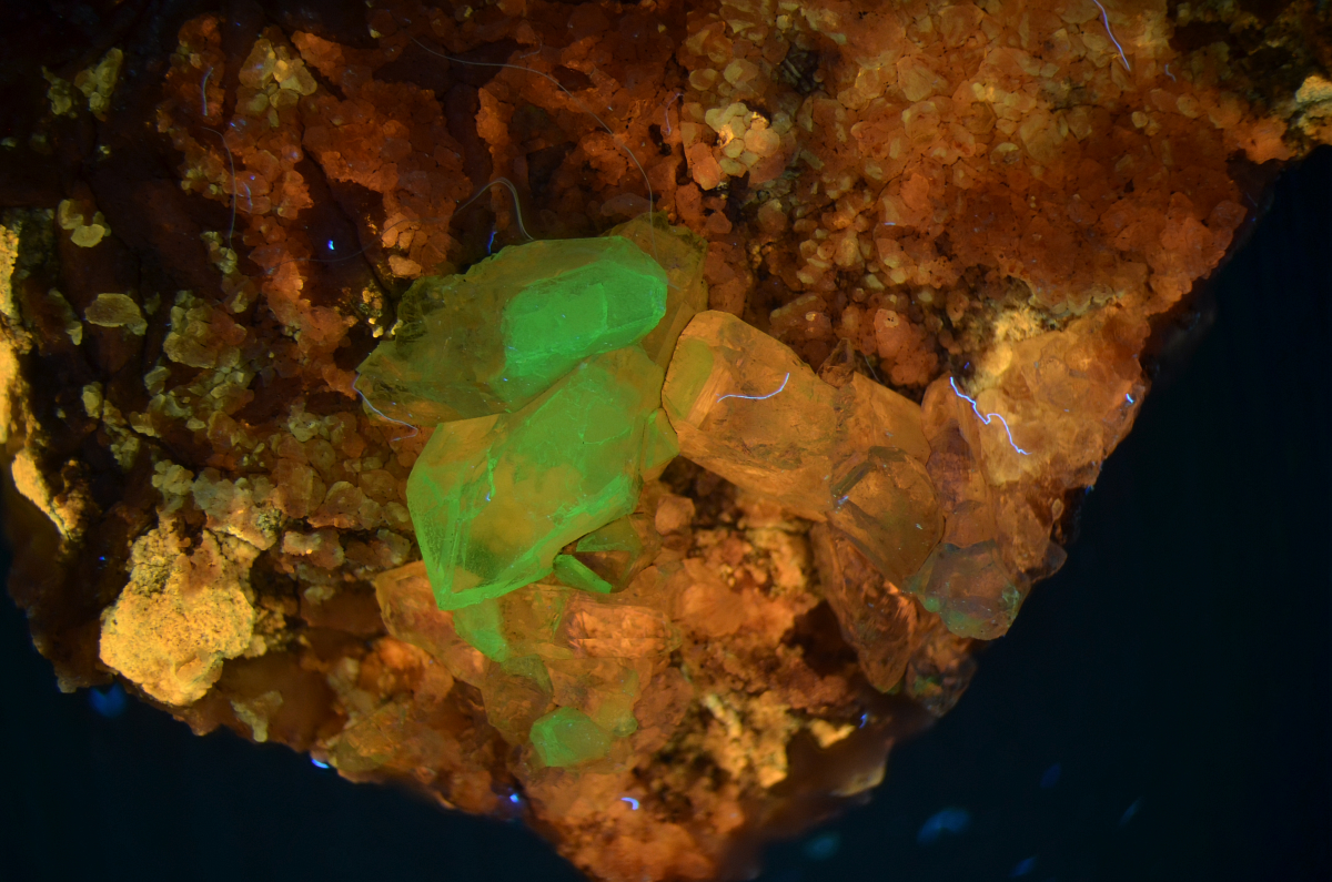
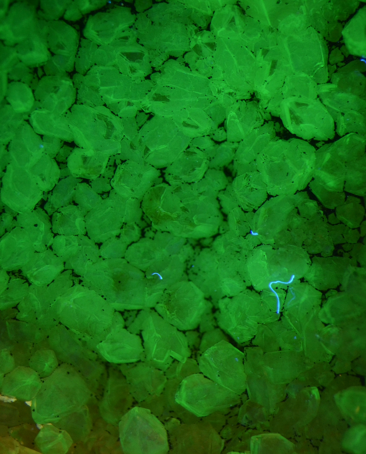
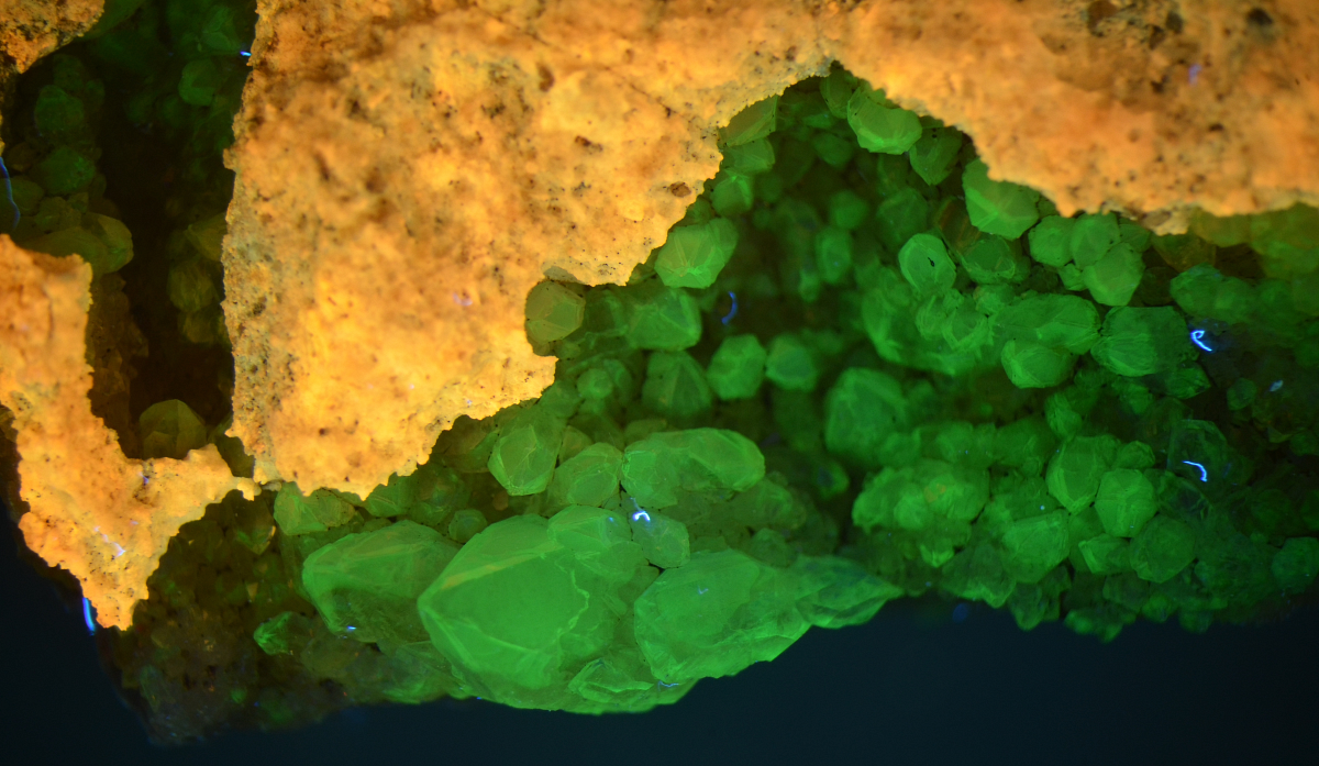
The quartz crystals below weathered out of a Paleozoic limestone. The limestone has been partially recrystallized to a marble. So far, we have found only a small number of these fluorescing quartz crystals in the float, none in outcrop. The activator is likely uranium and appears to be restricted to a thin overgrowth on the otherwise clear quartz crystals.
Fluorite!
These fluorite octohedra occur in outcrop in a vein filling with goethite included druzy quartz. Host rock is Paleozoic limestone.
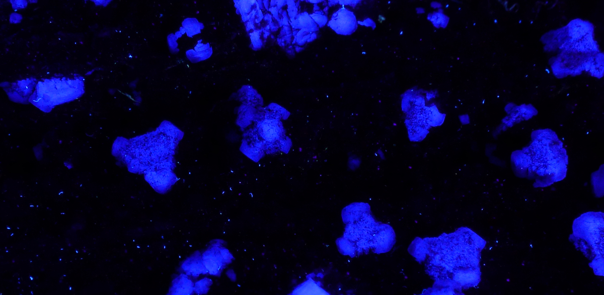
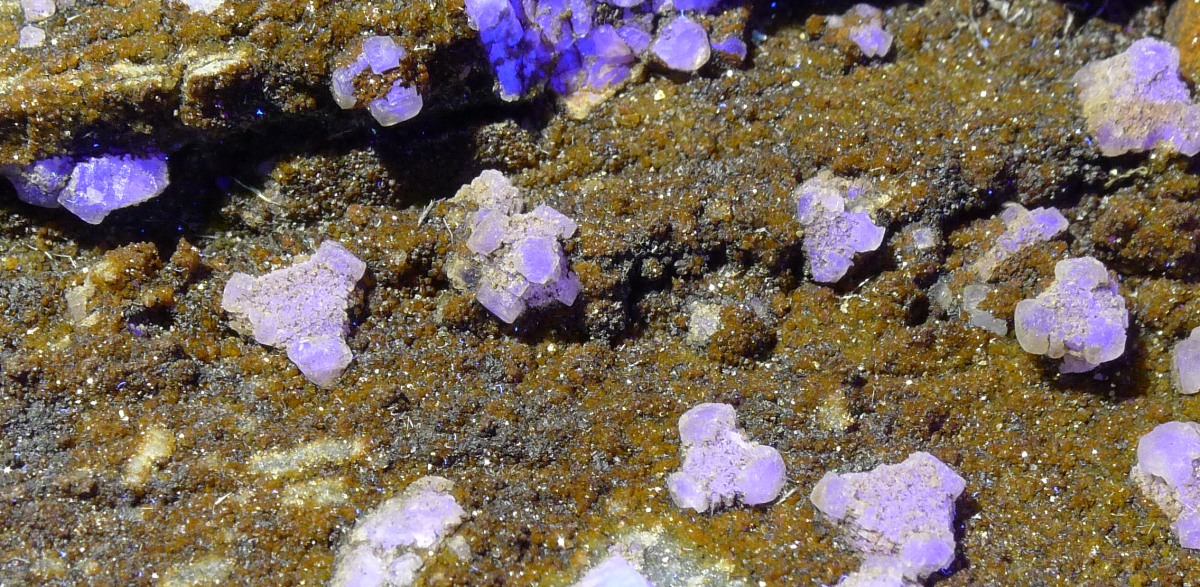
From the same locality as the octohedra above, these specimens show a brecciated limestone, likely fault gouge ,with fluorite veins which were themselves brecciated and then infilled with calcite veins. The calcite veins show orange under shortwave UV and white or pale yellow under longwave UV. Yellow caliche is more recent and likely formed post-mining.
Scheelite!
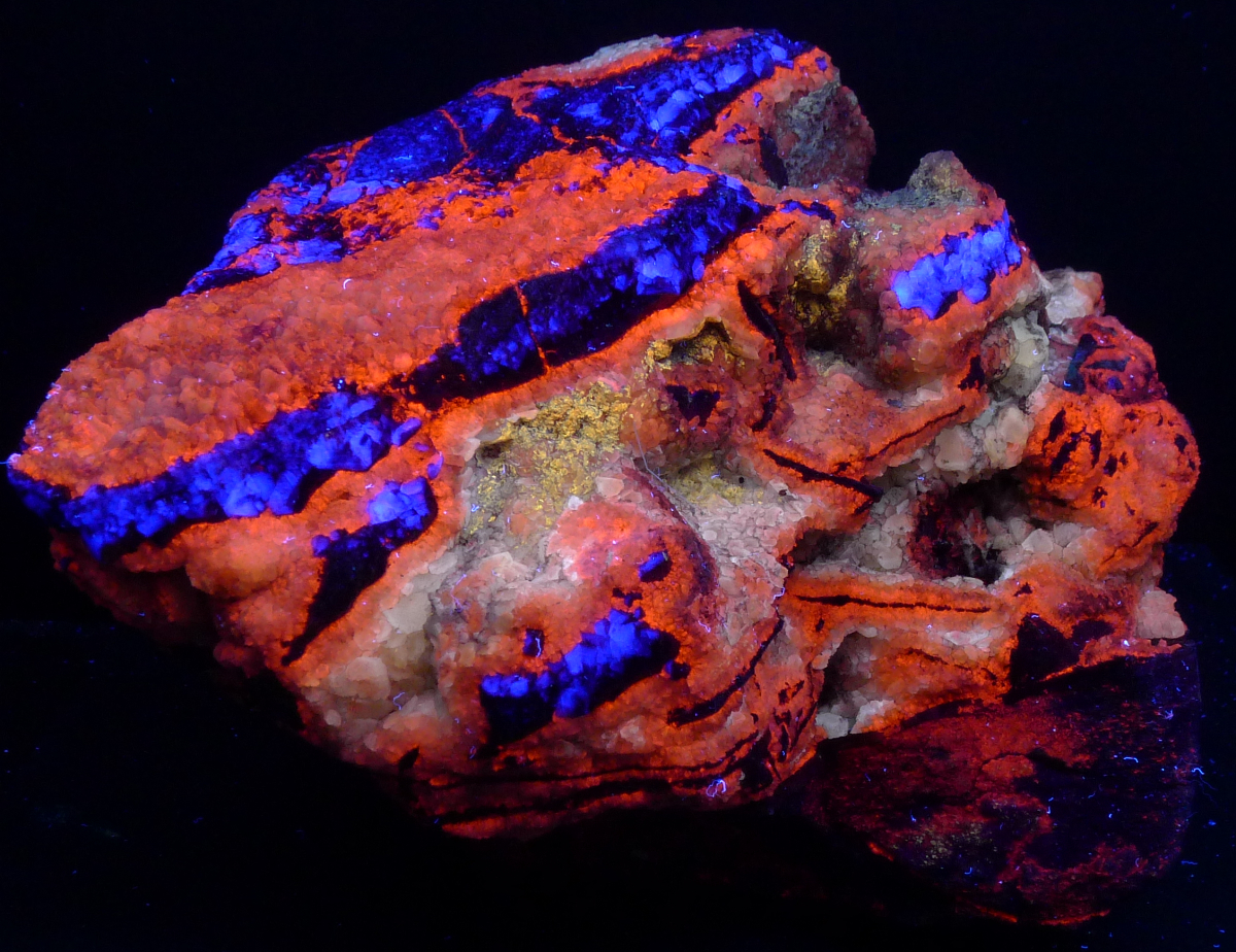
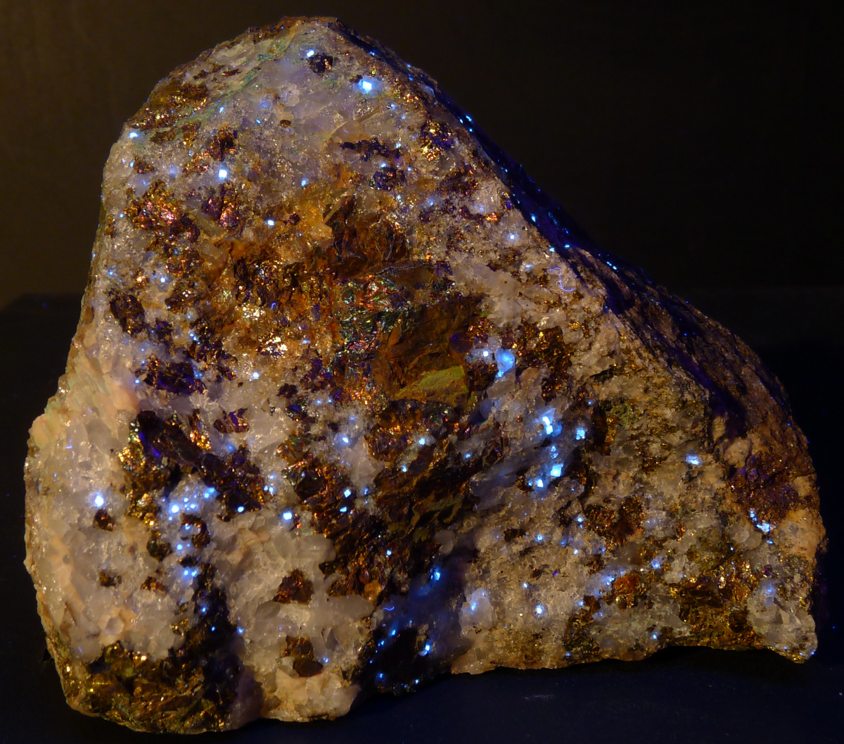
Quartz and bornite specimen with accessory calcite and scheelite from skarn hosted in Paleozoic limestone and quartzite.
Notes on Photography
The photos on this page were taken using Nikon D90, D5100, Lumix LX5, and Sony RX100 cameras. Exposure times range from 3 to 60 seconds. See the exif data for each photo for more information on exposures. For some of the photos minor sharpening, lightening, and contrast enhancement was applied to render the images as true to the naked eye appearance of the specimens as possible. A variety of white balance settings were employed. Of the white balance methods tested, using a color temperature of 7000K was found to produce images with the closest match to the naked eye. Thin section photomicrographs were taken under an Olympus-Elgeet POS petrographic microscope with a Nikon D90. Quartz crystal photomicrographs were taken under a LOMO MBC-10 stereo microscope with a Nikon D5100. All photos taken using a Way Too Cool 9 watt dual shortwave/longwave ultraviolet lamp and/or a MinerShop 18 watt shortwave lamp.
All images ©2012, 2013, 2014, 2015, 2016 Daniel Moore